On October 15, the hospital announced that the patient was T.T.Q.N, a 10-year-old from Dong Nai province, who was taken to Children 2 Hospital due to prolonged cough and shortness of breath. Through a CT scan, doctors discovered a massive lung tumor invading the entire left lung, simultaneously causing bronchial obstruction at the left and left pulmonary arteries, forcing N. to breathe with only the right lung.
Judging it a very difficult and dangerous operation, the surgical team decided to carry out a needle biopsy with the hope that if it was a malignant tumor, chemotherapy would help reduce its size, thus helping the surgery to be more feasible and safer. However, the biopsy revealed that it was a CPMT tumor.
MSc., Doctor Vu Truong Nhan, Deputy Head of the hospital’s Department of General Surgery, said that it was a very rare tumor and was bordering on malice that was not responsive to chemotherapy. According to the WHO document, by 2015, only 16 cases were reported worldwide and are usually found at neonatal stage due to prenatal diagnosis. But N. had no clear symptoms without antenatal diagnosis before, forcing her to carry the tumor for 10 years. Therefore, the tumor had quietly developed into a large invasion of the left lung. This may be the first case in the world recorded at this age with such large size.
Such difficult operation had discouraged many doctors, as the tumor was too large that had invaded the pericardial cavity, pulmonary veins and bronchi on the left, making surgery was very difficult and prolonged; only a minor mistake would lead to the death immediately on the operating table.
In addition, after the removal of the tumor, the pericardial cavity must be reconstructed to prevent cardiac herniation, a very serious complication. Moreover, postoperative complications for the left lung cut, including mediastinum oscillation, twist in bronchi and large blood vessels and scoliosis, although rare, may occur after the major surgery. In addition, the patient should be monitored closely every day after the major left lung cut. All these difficulties easily hinder the doctors.
However, due to the dangerous nature of the tumor, if the surgery was not performed, the tumor might block the arteries and bronchi, causing the sudden death. Against such danger and with the allowance from the child’s parents, the surgical team was determined to perform the operation to save N.
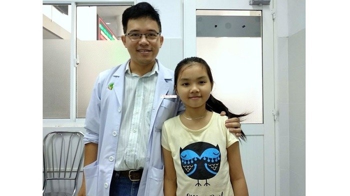
The surgical team had to subtly remove the tumor and it took them around eight hours of tension to completely eliminate the tumor weighing 1.5 kg. The patient had resiliently overcome all the challenges. The results of a six-month follow-up visit showed that the child has recovered completely and has no complications.


